Research Article - Onkologia i Radioterapia ( 2020) Volume 14, Issue 4
Aneurysmal bone cyst after femoral flexible elastic nailing: a case report
Alaa Jameel A. Albarakati1, Anfal J. Alsharif2, Daad F. Bin-Afif2, Linah A. Alhamdani2, Nada O. Hussain1, Roaa A. Saati2 and Ashraf Albrakati3*2College of Medicine,, Umm Alqura University, Makkah, Saudi Arabia
3Department of Human Anatomy, College of Medicine, Taif University, Taif, Saudi Arabia
Ashraf Albrakati, Department of Human Anatomy, College of Medicine, Taif University, Taif, Saudi Arabia, Email: Dr.albrakati@yahoo.com
Received: 19-Aug-2020 Accepted: 28-Aug-2020 Published: 15-Sep-2020
Abstract
Aneurysmal Bone Cyst (ABC) is a benign osteolytic blood-filled lesion but can be locally aggressive and potentially recurrent, mainly seen in paediatric population and young adults. ABC is thought to occur either due to certain gene mutations or secondary to a specific pathophysiologic change. We reported a case of a 5-year-old female patient presented with the left femur fracture that was treated with a flexible elastic nail fixation. Six months after the elastic nail was removed, she presented with left hip pain and limping. Radiography showed a cystic lesion in the proximal left femur, and the diagnosis of aneurysmal bone cyst was made. The lesion was treated with surgical curettage and bone graft. In conclusion, we would like to point out that aneurysmal bone cyst could develop secondary to the flexible elastic nail fixation. mp3download.link Best YouTube to MP3 converter. Download MP3 from YouTube for Free. one would honestly have expected more on the track mp3-go.net Download Mp3 songs for free Given the legendary pedigree of the man behind the sound downloadmp3-gratis.biz Download mp3 songs online at Mp3 Converter, watch high quality online music videos download-mp3gratis.me watch and download free songs of the highest quality. Listen to songs online here comfortably without any annoying advertisements. metrolagu.com Easy to use and free MP3 downloader. YouTube To MP3 download in seconds using the best YouTube to MP3 converter. YouTube Mp3 Get the latest song by simply typing the latest artist or song title in the Search menu. Mp3 file format with 128 - 320 Kbps bitrate converted from YouTube videos. read at this blog All those artist performances are still available on YouTube today find more here
Keywords
Aneurysmal bone, cyst, benign, trauma, osteolytic lesion
Introduction
ABC is a benign osteolytic blood-filled lesion. It generally involves metaphysis of the long bones and the vertebral column. Despite the benign nature, it can be locally aggressive and potentially recurrent [1]. It is usually present in the paediatric population and young adults with no sex predominance [2]. ABC’s are thought to be either as a result of certain gene mutations or secondary to a specific pathophysiologic change such as trauma, reactive vascular malformation, or surgical intervention [3]. The aim of this study was to present a case of an ABC developed after a flexible elastic nail fixation, which, to the best of our knowledge, has never been reported before.
Case Representation
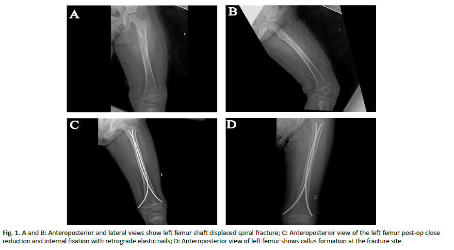
A 5-year-old female presented to the emergency room complaining about left thigh pain after a fall from 2 m height while playing. Her pain was continuous, and she could not walk. Past medical history was unremarkable. On the examination her left thigh was swollen, tender, and deformed. The neovascularity was intact. Radiography showed a left femur fracture (Figure 1A and 1B). The patient was admitted for surgery, closed reduction and internal fixation of the left femur with two intramedullary flexible titanium elastic nails was performed.
Figure 1: A and B: Anteroposterior and lateral views show left femur shaft displaced spiral fracture; C: Anteroposterior view of the left femur post-op close reduction and internal fixation with retrograde elastic nails; D: Anteroposterior view of left femur shows callus formation at the fracture site
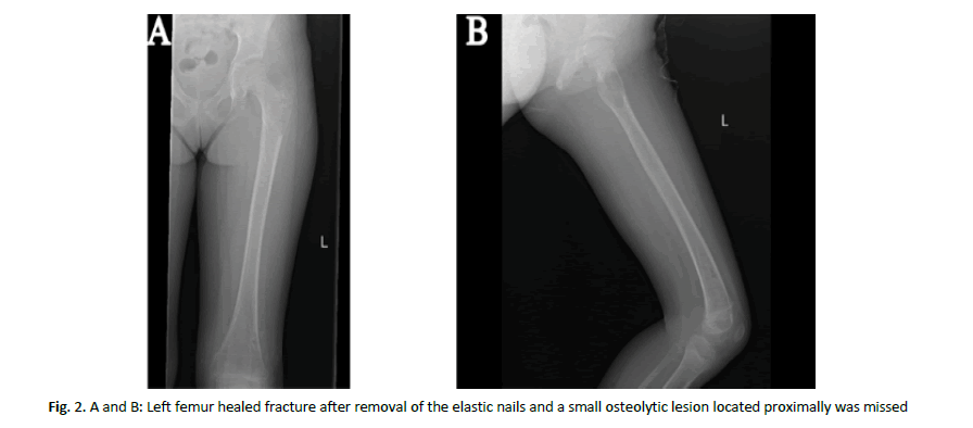
Two weeks later, the sutures were removed, and the patient avoided weight bearing on the left limb. After six weeks, during an ambulatory visit, no tenderness was found, but she was afraid of walking, and her x-ray film revealed good callus formation (Figure 1C). She was referred to the physiotherapy, where she started to improve her ability to walk. The patient was examined again 6 months later, she was without complains and walking normally with full range of motion in both hips and knees, the radiographs are shown in (Figure 1D). She was admitted for surgical removal of the elastic nails after 9 months. After the surgery, our patient had returned to her normal activity with full weight bearing on both limbs and no complications. The x-ray film after the removal of the elastic nails showed healed fracture but the specialist had missed a small osteolytic lesion in the proximal femur (Figure 2A and 2B).
Figure 2: A and B: Left femur healed fracture after removal of the elastic nails and a small osteolytic lesion located proximally was missed
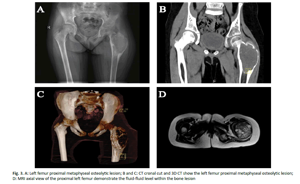
Consequently, six months after removing the flexible elastic nail, she presented to the clinic with left hip pain and limping for one month. The pain was continuous and aggravated with standing and walking. On the examination, her vital signs were stable, and she was had antalgic gait. No visual deformity, skin changes, and leg length discrepancies were observed. She had a tender palpable mass over the left hip. We also observed limited abduction of the left hip joint, but it was neurovascularly intact. Her radiological images showed a metaphyseal, eccentric, expansile, aggressive, and osteolytic bone lesion observed exactly where was the lateral nail tip located proximally. It had well defined margins along with a smooth inner margin and a rim of bone sclerosis with thin cortex. However, no periosteal reaction observed (Figure 3A-3C).
Magnetic resonance imaging revealed multil ocular cystic lesion measured 8.5 × 6.5 × 5.5 cm in maximum craniocaudal, anteroposterior, and transverse diameters, respectively. It showed a heterogeneous signal with hemorrhagic foci and fluid-fluid levels. The lesion was well defined, with narrow transition zone, associated with expansion, endosteal scalloping, and cortical thinning. We found limited edema of the adjacent soft tissue but no matrix mineralization, periosteal reaction, or soft tissue component (Figure 3D).
Figure 3: A: Left femur proximal metaphyseal osteolytic lesion; B and C: CT cronal cut and 3D CT show the left femur proximal metaphyseal osteolytic lesion; D: MRI axial view of the proximal left femur demonstrate the fluid-fluid level within the bone lesion
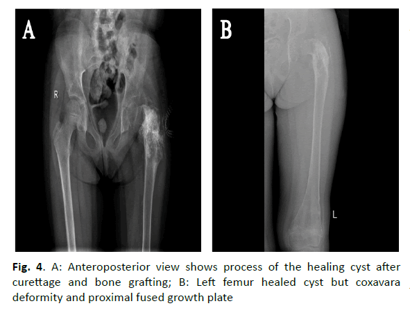
The patient underwent surgery with incisional biopsy, intralesional curettage, bone grafting, and application of the hip spica under general anaesthesia. The biopsy specimen was sectioned and stained with hematoxylin-eosin. The histopathological analysis showed a large cystic lesion filled with blood and separated by fibrous septa, alternating with solid areas. Clusters of osteoclasts like multinucleated giant cells with loose spindly to cellular stroma, reactive woven bone, and rare mitoses also were seen. No signs of malignancy were found. Diagnosis of ABC was confirmed. After 2 weeks, patient’s suture and hip spica were removed, but she kept non-weight bearing regime for 3 months. In her next visit, the patient presented to the clinic with no pain and x-ray films showed healed cyst; therefore, physiotherapy was initiated (Figure 4A).
Figure 4: A: Anteroposterior view shows process of the healing cyst after curettage and bone grafting; B: Left femur healed cyst but coxavara deformity and proximal fused growth plate
Unfortunately, 6 months later the patient presented with limping but no pain, her x-ray film showed coxa vara deformity of the left hip and completely healed lesion (Figure 4B). Finally, the patient was referred to the paediatric orthopaedic department for the correction of the deformity and further management.
Discussion
Our study purpose was to present that the flexible elastic nails could be the underlying cause of ABC. Regrettably, the knowledge is limited regarding the etiology, natural history, and treatment outcome of the ABC, despite the long experience in clinical practice [3]. Several studies have proposed different theories about the pathogenesis of ABC. One theory suggested that the lesion is a result of a vascular degenerative process due to other bone injuries, including giant cell tumour, osteoma, and other tumours, in genetically predisposed patients [4, 5].
However, the pathological findings did not support this proposal. While other studies showed that vascular malformation could be the source of ABC progression, by increasing the pressure on the bone, as in return aggravating the erosion and possibly leading to the resorption of the bone [4]. Additionally, a study published in 2007 claimed that etiological factors as trauma and surgical intervention may have a role in the pathogenesis of ABC. In that, the process of vascular alternation and remodelling of the injured tissues takes place [5]. In a case report of Dagher et al. a patient with ABC presented 5 years after the anterior cruciate ligament repair. Here, we cannot assume a causative relation between the surgical trauma and ABC formation, since the patient had a previous trauma that could be the cause of ABC [6]. Another case of the ABC, which occurred after the resection of an intraarticular nodular fasciitis of the elbow was described by Yamamoto et al., who had mentioned that ABC and nodular fasciitis have comparable genes. Surgical trauma should be considered as a part of the pathogenesis in this case [3]. Lastly, Sahin K et al. has reported a case of ABC after a bilateral femoral derotational osteotomy, which supports the theory that surgical intervention has a role in the pathogenesis of ABC [7]. Most ABC cases are classified as primary bone lesions, but the possibility of the bone lesions after the fixation of the flexible elastic nail should be considered, as reported in our case.
Conclusion
The study concluded that aneurysmal bone cyst could develop secondary to the flexible elastic nail fixation.
References
- Dorosh J, Vyas P. Aneurysmal bone cyst of the distal femoral metaphysis in a four-year-old female patient presenting with a pathologic fracture: a case report. Cureus. 2019;11:e4846.
- Park HY, Yang SK, Sheppard WL, Hegde V, Zoller SD, et al. Current management of aneurysmal bone cysts. Curr Rev Musculoskelet Med. 2016;9:435-444.
- Yamamoto M, Hiroshi U, Yoshihiro N, Hitoshi H. Secondary aneurysmal bone cyst in the distal humerus after resection of intra-articular nodular fasciitis of the elbow. BMC Res Notes. 2015;8:311.
- Mankin HJ, Francis J, Ortiz-Cruz HE , Villafuerte J , Gebhardt MC. Aneurysmal bone cyst: a review of 150 patients. J Clin Oncol. 2005;23:6756-6762.
- Cottalorda J, Bourelle S. Modern concepts of primary aneurysmal bone cyst. Arch Orthop Trauma Surg. 2007;127:105-114.
- Dagher AP, Magid D, Johnson CA, Mccarthy EF, Fishman EK. Aneurysmal bone cyst developing after anterior cruciate ligament tear and repair. AJR Am J Roentgenol. 1992;158:1289-1291.
- Sahin K, Demirel M, Turgut N, Arzu U, Polat G. Aneurysmal bone cyst after femoral derotational osteotomy: a case report. Malays Orthop J. 2019;13:54-48.


