Case Report - Onkologia i Radioterapia ( 2023) Volume 17, Issue 8
Angioleiomyoma of the urinary bladder and review of the literature
Xhevdet Cuni1, Sabit Mehmet1, Arber Neziri1, Leutrim Cuni1, Donat Cuni2 and Labinot Shahini3*2Faculty of Medicine, College "Rezonanca", Pristina, Kosovo
3Institute of Pathology, College "Rezonanca", Pristina, Kosovo
Labinot Shahini, Institute of Pathology, College "Rezonanca", Pristina, Kosovo, Email: labinot.shahini@uni-pr.edu
Received: 25-Jul-2023, Manuscript No. OAR-23-107397; Accepted: 20-Aug-2023, Pre QC No. OAR-23-107397 (PQ); Editor assigned: 27-Jul-2023, Pre QC No. OAR-23-107397 (PQ); Reviewed: 10-Aug-2023, QC No. OAR-23-107397(Q); Revised: 17-Aug-2023, Manuscript No. OAR-23-107397 (R); Published: 24-Aug-2023, DOI: -
Abstract
The case of a 39-year-old man who had irritative voiding symptoms presented with burning sensation, dysuria, is reported. Initial abdominal ultrasound demonstrated normal kidney aspects and one suspicious lesion of the urinary bladder. The mass detected within his urinary bladder by several imaging methods, cystoscopy, and CT scan by retrograde contrasted cystography. The histological findings of the surgical specimen confirmed diagnosis of angioleiomyoma of the urinary bladder. This benign tumor is rarely diagnosed in urinary tract and can be mistaken for urothelial carcinoma. The clinical presentation, imaging findings, and management of this relatively rare benign tumor are discussed. We retrospectively reviewed many articles published in the Europe, USA, and Asia using PubMed, Scopus, Medscape, Medline, and the several scientific journals.
casinoplus vdcasino betriyal betriyal betriyal betriyal betriyal betriyal betriyal betriyal betriyal casinoplus casinoplus casinoplus maltcasino almanbahis melbet betsat fenomenbet betmatik
Keywords
angioleiomyoma, neoplasm of urinary bladder, cystoscopy
Introduction
Angioleiomyomas or vascular leiomyoma are smooth muscle tumors, which arise from the tunica media (smooth muscle layer) of the subcutaneous blood vessels [1, 2]. It is also known as an angiomyoma, vascular leiomyoma or dermal angioma. Angioleiomyomas typically present as slow-growing masses and can be asymptomatic throughout in the body.
Angioleiomyoma located in the urethra and ureter has been reported by several authors [3-5] but localized in the urinary bladder has not been reported by any author. We can also find many articles related to another nearby benign tumours in urinary tract such as leiomyoma, which has been identified and located in the urinary bladder [6].
Angioleiomyomas are most frequently reported in middle-aged female patients, they can be found throughout the body in male and female adults of all ages [7]. The underlying cause of angioleiomyoma is still unclear. Although no pathophysiological mechanisms have been described to explain the occurrence of this tumours, it might be related to an endocrine alteration [8-9].
Case Report
The case of a 39-year-old man who had irritative voiding symptoms presented with burning sensation was reported as dysuria. Poor symptoms relief after several antibiotic treatments for urinary tract infections was documented. Laboratory analyzes of urine confirmed micro-hematuria. Initial abdominal ultrasound demonstrated normal kidney aspects and one well defined lesion in the urinary bladder about 2.5 cm in diameter. The mass was located in posterior wall of urinary bladder. Cystoscopy examination described one well localized intravesical lesion with extended pedicle. Based on cystocopy examination this lesion was initially suspected to be a urothelial carcinoma (TCC) and surgical intervention potentially, including TUR-B, was considered. After discussion with the patient, a decision was made to resect the mass by endourology TUR-BT procedure. The intervention was performed under spinal anaesthesia, but resection of the suspected mass was not achieved.
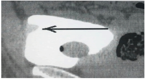
This lesion was non-resectable in TUR-BT and this was a surprising fact. We requested the examination by CT scan with contrasted retrograde cystography to classify the location of the suspicious lesion in relation to the urinary bladder wall (Figure 1). The CT scan examination confirms the intramural location of the lesion.

Figure 1: Ct scan image of the contrasted retrograde cystography
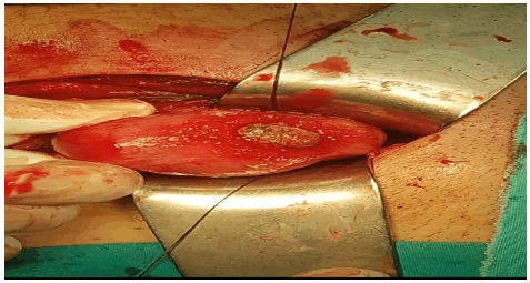
After the second discussion with the patient, a decision was made for the next operative procedure by open suprapubic incision under spinal anaesthesia. Intraoperatively by cistostomy the carefully excision en-bloc of this bladder lesion was done respecting the margins (Figure 2).

Figure 2: Surgical image from the operative wound
The sample has been sent in Institute of Pathology for histopathology examination.
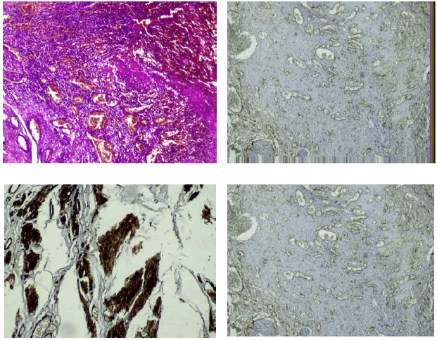
Immunohistochemistry showed significant histopathologic findings. Microscopically, areas of mixture of smooth muscle bundles arranged in small fascicles are visible and intervening vascular channels is noted (Figure 3). Dilated vascular channels, the walls of which are difficult to distinguish from the intervascular smooth muscle.

Figure 3: Intramural angioleiomyoma of urinary bladder-cavernous type
Discussion
Angioleiomyoma are commonly deep, well-circumscribed smooth muscle tumours that originate from the walls of blood vessels in particular veins. Although the neoplasm can affect any age, it is commonly seen in adults between the age of 30 and 60 years of age. Typically, the lesion presents as a solitary, small, slow growing, nodule [1, 2, 7].
Angioleiomyoma localized in the parts of the urinary tract are rare. Only few cases that arise in urethral and ureter location have been reported in literature [3-5].
Another type of benign tumour’s Leiomyomas localized in the parts of the urinary tract was reported by several authors [10-15].
Leiomyomas constitutes 35% of benign mesenchymal bladder tumours and most cases of urogenital leiomyoma have been described in Japanese publications [14]. Histologically angioleiomyomas are characterized as a round encapsulated lesions which are composed of many blood vessels of variably thickened walls that are surrounded by interlacing fascicles and bundles of uniform spindled cell. True angioleiomyomas belong to the family of Perivascular Epithelioid Cell Tumours (PEComas), however, angioleiomyoma do not show the characteristic HMB-45 positive staining of PEComas. Three histologic variants have been recognized; the most common one being “capillary or solid” type representing approximately 67% of cases. Other variants include “venous” and “cavernous” types. Identification of these histologic variants is important for pathologists to be able to identify them and recognize them as variants of the same benign entity [8, 9]. Immunohistochemistry shows cells to be positive for smooth muscle actin, calponin, and h-caldesmin. Surgical excision by TUR-BT is the recommended treatment for any intravesical lesion mainly to identify the lesion histologically. In our case it failed. The open operative treatment by cystotomy in our clinical conditions was the right operative decision. Recurrence of angioleiomyoma or any malignant lesion after simple excision in our case was not confirmed in follow-up by regular cystoscopy every 6 months in cohort period of 2 years. Based on the works of many authors, different treatment procedures for selected angioleiomyoma by complete surgical excision are the key to success and no recurrence was reported [15-17].
Conclusion
Intramural vesical angioleiomyomas are rare cases of benign tumor in the urinary bladder and that fact highlights the importance of thorough evaluation and histopathological examination in patients with suspicious lesion in urinary bladder. Although the angioleiomyomas of the urinary tract should be considered in the differential diagnosis of other more often diagnosed urothelial tumors. In general, cystoscopy and CT can be used to classify suspicious urinary vesical lesions by its locations into three positions: endovesical, intramural, or extravesical. Mostly of benign bladder urinary tumors are result of the submucosal growth of the leiomyoma, and were first described by Campbell and Gislason; they are usually polypoid or pedunculated. The small intramural tumors are reported as a less symptomatic. A histopathological analysis is necessary for an accurate diagnosis and appropriately selected management. So far they have not been reported in the literature about recurrences or malignant degenerations of leiomyomas in the urinary bladder and angioleiomyomas in urethra. Treatment of the urinary bladder tumours (benign and malign) is surgical.
TUR-BT it still counts as an actual treatment procedure in the solution of selected small bladder tumours.
In general, in selected cases where bladder tumours have progressed in growth applying the assessment according to TNM staging is very important. The segmental resection or partial cystectomy should be considered. Surgical excision has excellent prognosis after complete resection and should always be offered.
We reported the case of angioleiomyomas localized in the urinary bladder wall, successfully removed by open surgical operationcystectomy without recurrence and without compromising the bladder capacity.
Acknowledgment
There is no conflict of interest or financial acknowledgments for this case report.
References
- Heffernan MP, Smoller BR, Kohler S. Cutaneous epithelioid angioleiomyoma. Am J Dermatopathol. 1998;20:213-217.
- Requena L, Baran R. Digital angioleiomyoma: an uncommon neoplasm. J Am Acad Dermatol. 1993;29:1043-1044.
- Monzon J., Rodriguez G., Trilla S. Obstructive urethral angileiomyoma. Arch Esp Urol. 2015; 57:1128-1130.
- Pacik D, Dolezel J, Skoumal R, Bucek J, Kladenský J. Very rare angioleiomyoma of the male urethra. Int Urol Nephrol. 1993;25:479-484.
- Krivoborodov GG, Raksha AP, Malenko VP. Angioleiomyoma of the ureter. Urologiia. 2001;1:43-45.
- Campbell EW, Gislason GJ. Benign mesothelial tumors of the urinary bladder: review of literature and a report of a case of leiomyoma. J Urol. 1953; 70:733-742.
- Murata H, Matsui T, Horie N, Sakabe T, Konishi E, Kubo T. Angioleiomyoma with calcification of the heel: report of two cases. Foot Ankle Int. 2007; 28:1021-105.
- Amir RA, Sheikh SS. Angioleiomyoma of urethra: A case report. Int J Surg Case Rep. 2015; 10:195-197.
- Phillip H.. McKee, Calonje E, Scott R.. Granter. Pathology of the Skin: With Clinical Correlations. Elsevier Mosby; 2005.
- Kato T, Kobayashi T, Ikeda R, Nakamura T, Akakura K, Hikage T, Inoue T. Urethral leiomyoma expressing estrogen receptors. Int J Urol. 2004; 11:573-575.
- Cheng C, Lai FM, Chan PS. Leiomyoma of the female urethra: a case report and review. J Urol. 1992;148:1526-1527.
- O'Connell K, Edson M. Leiomyoma of bladder. Urology. 1975; 6:114-115.
- Katz RB, Waldbaum RS. Benign mesothelial tumor of bladder. Urology. 1975; 5:236-238.
- Nishiyama H, Nakamura K, Nishimura M, Nishimura K, Takahashi Y. Male bladder leiomyoma: a case report. Hinyokika kiyo. Acta Urologica Japonica. 1992; 38:949-952.
- Hachisuga T, Hashimoto H, Enjoji M. Angioleiomyoma. A clinicopathologic reappraisal of 562 cases. Cancer. 1984; 54:126-130.
- Amir RA, Sheikh SS. Angioleiomyoma of urethra: A case report. Int J Surg Case Rep. 2015; 10:195-197.
- Khater N, Sakr G. Bladder leiomyoma: Presentation, evaluation and treatment. Arab J Urol. 2013; 11: 54-61.


