Case Report - Onkologia i Radioterapia ( 2023) Volume 17, Issue 2
Management of infected Urachal Cyst and the pattern of drug resistance in the isolate
Priti Singh1, Salman Khan2* and Shakeel Ahmed22Department of Pathological Processes and Therapeutics, American University School of Medicine Aruba, Aruba
Salman Khan, Department of Pathological Processes and Therapeutics, American University School of Medicine Aruba, Aruba, Email: salman186631@gmail.com
Received: 16-Feb-2023, Manuscript No. OAR-22-89522; Accepted: 11-Mar-2023, Pre QC No. OAR-22-89522 (PQ); Editor assigned: 18-Feb-2023, Pre QC No. OAR-22-89522 (PQ); Reviewed: 03-Mar-2023, QC No. OAR-22-89522 (Q); Revised: 10-Mar-2023, Manuscript No. OAR-22-89522 (R); Published: 15-Mar-2023
Abstract
It is defective obliteration of the urachus that causes urachal cysts. Failure of urachus to involute can contribute to anomalies that may increase the risk of infection and/or malignancy if left untreated.
A 29-year-old female with clinical features suggestive of acute abdominal pain is presented in this case report. Complete blood counts, serum electrolytes, urinalysis, renal function tests were normal. In the suprapubic region, an ultrasonographic examination revealed a hypoechoic lesion measuring 2 cm × 1.5 cm in size and contrast-enhanced CT scan confirmed a non-communicating urachal cyst between abdominal wall and urinary bladder's anterior wall.
Urine culture revealed significant growth of Escherichia coli and was susceptible to eight out of fifteen antibiotics. The patient was successfully treated with parenteral amikacin.
In clinical office, urachal cysts need to be diagnosed and managed promptly with a high degree of suspicion. Radiology, urine culture & sensitivity are effective strategies to curb the acute presentation of Urachal Cyst.
Keywords
urachus, urachal cyst, sepsis, Escherichia coli, acute abdomen, Radiology, amikacin
Introduction
The urachus is the remnant of allantois inside the abdomen. This duct connects fatal bladder with umbilicus [1]. Later it gradually obliterates and becomes a fibrous tract [2].
Various anomalies of urachus may appear due to defective or incomplete obliteration, such as patency, sinus, diverticulum, or cyst formation [3]. The cyst is one of the most common anomalies and develops when a segment of the urachus obliterate leaving a cystic cavity [4].
Pathologically transitional epithelium forms the inner, connective tissue middle, and smooth muscle fibres the outermost layer of the urachal cyst [5]. Normally it remains asymptomatic, until it gets infected [6]. Following an infection, it presents as a painful mass below umbilicus [7]. Infected urachal cyst may present with other signs and symptoms such as lower abdominal pain, difficulty in urination, and a bloody or purulent discharge from the umbilicus [8].
This infection and acute abdomen picture often lead to multiple differentials therefore; a physician should be highly suspicious to reach the accurate diagnosis. Sometimes the diagnosis is confused with incarcerated umbilical hernia, Inflammations of the appendix, Meckel diverticulum, urinary tract, and bladder cancer especially in smokers [9].
Diagnosis of urachal cyst needs timely investigations including ultrasonography, computed tomography and MRI. The ultrasound may reveal a hypoechoic lesion behind the abdominal wall. Similar lesion can be seen on CT scan around the urinary bladder. Imaging may discover other anomalies like urachal sinuses. These investigations are important preoperatively to define the size, vascularity, and proximity with the surrounding organs [10].
Needle aspiration with culture and sensitivity of the fluid is recommended for an appropriate antibiotic. Common infecting organisms are Escherichia coli, Enterococcus faecium, and Klebsiella pneumoniae [11].
The urachal cyst is managed on its pathology, chronicity, and presentation. A two-step procedure can be performed. Initially an acute fluctuant cyst can be incised and drain under an antibiotic cover. In the next step the remnant of the cyst can be excised completely. In subacute cases both the procedures may be combined [12]. The common surgical complications are infections, bleeding, and fistula formation [13].
In this report, we describe an acutely presented urachal cyst in a patient, which was accurately diagnosed and successfully treated by an appropriate antibiotic.
Case Presentation
A young woman came in emergency with complain of recurrent pain in the abdomen mostly the suprapubic area for about a week. There was no radiation or migration to other areas of the abdomen. She often complained about the difficulty in passing urine. No haematuria was reported. The patient denied any history of vomiting, jaundice and steatorrhea. Later the patient became febrile. On physical examination, the patient was an active woman with a good build and BMI. Her heart rate was 86 per minute, BP 110/76 mm of Hg, and temperature 38.0°C. On examination the abdomen was soft with a 3 cm sized tender swelling above the bladder. The blood counts, urinalysis, electrolytes, and renal function tests were found normal. Ultrasonography revealed a 2 cm × 15 cm sized lesion above the bladder. A Contrast-Enhanced CT scan (CECT) was done that confirmed a non-communicating urachal cyst between urinary bladder and anterior abdominal wall. The E. coli were grown in urine culture. The patient’s infection was successfully treated with parenteral amikacin.
Investigations
Urinalysis and renal function tests
Renal function panel and urinalysis were analysed which were found within normal range.
Urine Culture and Sensitivity
In our patient urinalysis did not reveal any pyuria, however E. coli was found by urine culture.
The sensitivity was tested against various antibacterial agents. The E. coli was found sensitive to eight out of fifteen antibiotics.
In our study the carbapenem was the lowest <=0.25 MIC while the penicillin and cephalosporin classes were among the most resistant >=16 MIC as shown in the Table 1.
Tab. 1. Resistance of Escherichia coli isolated from Urine culture to a panel of fifteen antibiotics
| S. No. | Antibiotics | MIC | Interpretation |
|---|---|---|---|
| 1 | Amikacin | <=4 | Sensitive |
| 2 | Ampicillin | <16 | Resistant |
| 3 | Aztreonam | 16 | Resistant |
| 4 | Cefazolin | <16 | Resistant |
| 5 | Cefepime | 4 | Sensitive |
| 6 | Cefoxitin | <=4 | Sensitive |
| 7 | Ceftazidime | 8 | Intermediate |
| 8 | Ceftriaxone | >4 | Resistant |
| 9 | Ciprofloxacin | >2 | Resistant |
| 10 | Colistin | <=1 | Sensitive |
| 11 | Ertapenem | <=0.25 | Sensitive |
| 12 | Fosfomycin | <=16 | Sensitive |
| 13 | Gentamicin | <=2 | Sensitive |
| 14 | Imipenem | <=0.25 | Sensitive |
| 15 | Levofloxacin | >4 | Resistant |
Plain X-ray KUB
In our patient no radio-opaque calculus or abnormal calcifications were found on plain scan, KUB.
Ultrasonography
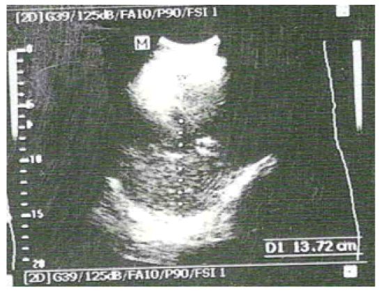
The bladder ultrasound of our patient revealed a cystic mass of 15mm × 11 mm size at urinary bladder's anterior wall (Figure 1).

Figure 1:Abdominal ultra-sonographic finding of a urachal cyst (longitudinal view)
Contrast-Enhanced CT (CECT)
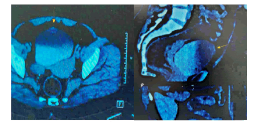
A Contrast-Enhanced CT scan (CECT) was done for our patient that shows a non-communicating urachal cyst between urinary bladder and anterior abdominal wall.
The kidneys and urinary tracts were normal in shape, size, and position. The pyelogram was found functionally within normal limits.
An ovoid cystic lesion measuring 24 × 20 × 14 with peripheral enhancement is seen at urinary bladder's anterior wall. No solid mass is seen (Figure 2).

Figure 2: . A contrast-enhanced CT scan (CECT) shows an ovoid cystic lesion with peripheral enhancement on the anterior wall of the urinary bladder.
Discussion and Conclusion
The urachus is a remanent of allantois (an embryological tube connecting the umbilicus and the bladder) [14].
During the latter part of fatal life, it gradually obliterates and becomes median umbilical ligament [15].
If there is a failure to obliterate, it may result in a patent urachus, cyst or fistula. Later due to pathological complications they may turn infected, inflamed or malignant [16].
Urachal remnant anomalies are less prevalent in population, about 2 in 1000. They are more commonly seen in boys. The prevalence of urachal cysts is reported as 1 in 600 [17].
The acute abdomen picture may result in diagnostic confusion. A suspicion should always be there in a presentation of acute abdomen to diagnose and manage a relatively rare condition. Our patient presented with a subacute picture leading to various other possibilities like hernia, appendicitis, ectopic pregnancy, diverticulitis, urinary tract infection and pelvic inflammatory disease [18].
The urinalysis report in our patient was found within normal limit. Although the statistical data shows evidence of infection in the bladder and urinary tract in an infected urachal cyst. It may reflect some active sediments including haematuria [19].
The urachal c ysts are often associated with lower abdominal tenderness, fever, dysuria, and genital infections, especially once they are infected [20]
The perforation has been reported by Campanale et al. leading to peritonitis and an urgent need for surgery [21].
In our patient urinalysis did not reveal any pyuria, however E. coli was found by urine culture.
The sensitivity was tested against various antibacterial agents. The E. coli was found sensitive to eight out of fifteen antibiotics. The Minimum Inhibitory Concentration (MIC) provides a physician valuable information for making a prescription. In our study the carbapenem was the lowest <=0.25 while the penicillin and cephalosporin classes were among the most resistant >=16 [22]
The sonography, CT scan, Contrast studies, and MRI are primary investigations to determine the size, inflammatory changes, and anatomical location of the urachal cyst. The b ladder u ltrasound of our patient revealed a cystic mass of 15 mm × 11 mm size anterior to the urinary bladder. A similar interesting ultrasound was reported by Yoo KH and Nagasaki A et al [23,24]
The focused imaging modalities are important to diagnose a lesion. CT scan remains the primary modality of choice specially to localize a small pathology. It differentiates likelihood of benign and malignant lesion. The goal of imaging is to differentiate between a solid and cystic lesion. Another help may be found to figure out any signs of pathological aggressiveness like benign or malignant changes. We have selected Contrast Enhanced CT Scan (CECT) as they focus on the lesions, not easily diagnosed on KUB, ultrasound or plain CT [25]
A Contrast-Enhanced CT scan (CECT) was done for our patient that shows a non-communicating urachal cyst between urinary bladder and the abdominal wall. The kidneys and urinary tracts were normal in shape, size, and position. found functionally within normal limits. An ovoid cystic lesion measuring 24 × 20 × 14 with peripheral enhancement is seen at the anterior wall of urinary bladder. No solid mass is seen. Ultrasonography Computed Tomography (CT) and contrastenhanced CT scan are the most common diagnostic imaging studies for any urachal remnant disease.
The urachal cyst is demonstrated at CECT as a sub-abdominal cystic lesion between the bladder and the umbilicus [26].
Tazi et al have described a 35-year-old Arab patient who presented with an acute abdomen in ER. The baseline investigations reflect a normal Urinalysis without significance bacteriuria. Th e er ect abdominal x-ray was normal. Ultrasonography of abdomen revealed some fluid in the peritoneal cavity. A helical CT abdomen showed inflammatory changes with an abscess formation between umbilicus and bladder, a further evaluation with an excretory time shows a small fluid collection above the bladder [27].
Nagasaki A et al. mentioned that Ultrasonography is an initial and cost-effective modality for diagnosing urachal cysts to rule in or out the suspected cyst. In a female patient sonography is always preferred over CT scan especially during pregnancy [28].
Urinalysis ruled out the possibility of urinary tract infection however urine culture showed the E. coli infection. The ultrasound ruled in favour of some cystic lesion. The diagnosis was confirmed by CECT. The imaging modalities of choice in urachal cyst remain sonography and CT scan [29].
Urachal cyst and associated urinary tract infections are commonly seen in children and adults. Our patient’s signs and symptoms were pertained to the infection and inflammation caused by Escherichia coli. The timely use of specific antibiotic cured the infection and abrogate a need for surgical intervention [30].
The medical management of the infection is important prior to the surgical intervention.
Newman et al. advised an earlier detection of infection and accurate antimicrobial management prior to surgery. It allows the surgical procedure safe and with minimum risks of complication. He reported that S. aureus is the most common organism found in the infected cyst [31].
The infected urachal cyst is traditionally managed by an antibiotic followed by a surgical excision [32].
Kibret et al reported E. coli isolation, mostly from the urine samples. The organism was highest sensitive to nitrofurantoin followed by other antimicrobials like, norfloxacin, gentamicin and ciprofloxacin (p<0.001) [33].
The most severe infections with multidrug-resistant were treated by aminoglycosides like amikacin. The common isolates were, Pseudomonas, Acinetobacter, Enterobacter, E. coli, Proteus, Klebsiella , and Serratia [34].
In our study the amikacin reflected MIC against E. coli <= 4. The treatment of our patient with injectable amikacin 750 mgs I/M × 5 days successfully improved the patient condition and later the patient was referred for a surgical consultation.
Author Contributions
Investigation, treatment, and resources by Dr. Priti Singh − “Conceptualization, Software, Dr Salman Khan, Writingoriginal draft preparation by Dr. Shakeel Ahmed.; writingreview and editing, Dr Salman Khan; Project administration, Dr Salman khan; All authors have read and agreed to the published version of the manuscript.
Statement Of Conflict Of Interest
Members of the committee declare that they have no conflict of interest
Reference
- Garcia LG, Ballesta B, Talavera JR, Orribo NM, Carrion A. Urachal pathology: review of cases. Urolog intern. 2022; 106:195-198.
- Choi YJ, Kim JM, Ahn SY, Oh JT, Han SW, et al. Urachal anomalies in children: a single center experience. Yons Med J. 2006; 47:782-786.
- Spina P, Chiari G, Minniti S. Intraperitoneal rupture of an infected urachal cyst: an unusual cause of acute abdomen in children. A case report and review of the literature. J Pediat Urol. 2006; 2:480-482.
- Sherman JM, Rocker J, Rakovchik E. Her belly button is leaking: a case of patent urachus. Ped Emerg Care. 2015; 31:202-204.
- Qureshi K, Maskell D, McMillan C, Wijewardena C. An infected urachal cyst presenting as an acute abdomen–A case report. Internat J Surgery Case Reports. 2013;4:633-635.
- Jayakumar S, Darlington D. Acute presentation of urachal cyst: a case report. Cureus. 2020;12.
- Urachal pathology: review of cases.
- Faye PM, Gueye ML, Thiam O, Niasse A, Ndong A, et al. Infected urachal cyst in an adult, report of two observations. Intern J Surgery Case Reports. 2022; 97:107-394.
- Ash A, Gujral R, Raio C. Infected urachal cyst initially misdiagnosed as an incarcerated umbilical hernia. J Emerg Med. 2012; 42:171-173.
- Yu JS, Kim KW, Lee HJ, Lee YJ, Yoon CS, et al. Urachal remnant diseases: spectrum of CT and US findings. Radiographics. 2001; 21:451-461.
- Kaya S, Bacanakgıl BH, Soyman Z, Kerımova R, Battal Havare S, et al. An infected urachal cyst in an adult woman. Case Reports Obstet Gynecol. 2015;201-205.
- Elkbuli A, Kinslow K, Ehrhardt Jr JD, et al. Surgical management for an infected urachal cyst in an adult: Case report and literature review. Intern J Surgery Case Reports. 2019; 57:130-133.
- Ashley RA, Inman BA, Routh JC, Rohlinger AL, Husmann DA, et al. Urachal anomalies: a longitudinal study of urachal remnants in children and adults. T J Urol. 2007; 178:1615-1618.
- Larsen MH, Hviid TV. Human leukocyte antigen-G polymorphism in relation to expression, function, and disease. Human immunology. 2009; 70:1026-1034.
- Choi YJ, Kim JM, Ahn SY, Oh JT, Han SW, et al. Urachal anomalies in children: a single center experience. Yon Med J. 2006; 47:782-786.
- Sherman JM, Rocker J, Rakovchik E. Her belly button is leaking: a case of patent urachus. Pediatric Emergency Care. 2015; 31:202-204.
- Keceli AM, Donmez MÄ°. Are urachal remnants really rare in children? An observational study. European J Pediatrics. 2021; 180:1987-1990.
- Ash A, Gujral R, Raio C. Infected urachal cyst initially misdiagnosed as an incarcerated umbilical hernia. The J Emerg Med. 2012; 42:171-173.
- Ozbek SS, Pourbagher MA, Pourbagher A. Urachal remnants in asymptomatic children: Grayâ?scale and color Doppler sonographic findings. Journal of Clinical Ultrasound. 2001; 29:218-222.
- Goldman IL, Caldamone AA, Gauderer M, Hampel N, Wesselhoeft CW. Infected urachal cysts: a review of 10 cases. J Urol. 1988; 140:375-378.
- Campanale RP, Krumbach RW. Intraperitoneal perforation of infected urachal cyst. Amer J Surg. 1955; 90:143-145.
- Poirel L, Madec JY, Lupo A, Schink AK, Kieffer N, et al. Antimicrobial resistance in Escherichia coli. Microbiology Spectrum. 2018; 6:6-14.
- Yoo KH, Lee SJ, Chang SG. Treatment of infected urachal cysts. Yonsei Medical Journal. 2006; 47:423-427.
- Nagasaki A, Handa N, Kawanami T. Diagnosis of urachal anomalies in infancy and childhood by contrast fistulography, ultrasound and CT. Pediatric Radiology. 1999; 21:321-323.
- Elstob A, Gonsalves M, Patel U. Diagnostic modalities. International Journal of Surgery. 2016; 36:504-512.
- Yu JS, Kim KW, Lee HJ, Lee YJ, Yoon CS, et al. Urachal remnant diseases: spectrum of CT and US findings. Radiographics. 2001; 21:451-461.
- Tazi F, Ahsaini M, Khalouk A, et al.Abscess of urachal remnants presenting with acute abdomen: a case series. J Med Case Reports.2012;6:1-7.
- Patel SJ, Reede DL, Katz DS, et al. Imaging the pregnant patient for nonobstetric conditions: algorithms and radiation dose considerations. Radiographics. 2007; 27:1705-1722.
- Daly KM, Rhouma SB, Zaghbib S, Oueslati A, Gharbi M, et al. Infected urachal cyst in an adult: a case report. Urology Case Reports. 2019; 26:100976.
- Elkbuli A, Kinslow K, Ehrhardt JD, Hai S, McKenney M, et al. Surgical management for an infected urachal cyst in an adult: case report and literature review. Int J Surg Case Rep. 2019,57:130-133.
- Newman BM, Karp MP, Jewett TC, Cooney DR. Advances in the management of infected urachal cysts. Journal of pediatric surgery.1986;21:1051-1054.
- Lipskar AM, Glick RD, Rosen NG, Layliev J, Hong AR, et al. Nonoperative management of symptomatic urachal anomalies. J Ped Surgery. 2010;45:1016-1019.
- Kibret M, Abera B. Antimicrobial susceptibility patterns of E. coli from clinical sources in northeast Ethiopia. African Health Sciences. 2011; 11:40-45.
- "Amikacin sulfate injection, solution". DailyMed. 2019.


