Case Report - Onkologia i Radioterapia ( 2023) Volume 17, Issue 11
Use of ultrasound as a primary imaging modality for screening adrenal metastasis in lung carcinoma patients-A case series in Indian population
Saranya Ravi*, Ayush Dixit, Sulabh Devaliya and Avadesh Pratap KushwahSaranya Ravi, Department of Radiodiagnosis, Netaji Subhash Chandia Bose Medical College and Hospital, Jabalpur, India, Tel: 9840793373, Email: saranyaraviiyer96@gmail.com
Received: 17-Mar-2023, Manuscript No. OAR-22-92079; , Pre QC No. OAR-22-92079 (PQ); Editor assigned: 21-Mar-2023, Pre QC No. OAR-22-92079 (PQ); Reviewed: 05-Apr-2023, QC No. OAR-22-92079; Revised: 17-May-2023, Manuscript No. OAR-22-92079 (R); Published: 25-May-2023
Abstract
Adrenal glands are paired endocrine glands located over the upper renal poles. Adrenal pathologies have various clinical presentations. They can coexist with the hyper function of individual cortical zones or the medulla, insufficiency of the adrenal cortex or retained normal hormonal function. The most common adrenal masses are tumors incidentally detected in imaging examinations (ultrasound, tomography, magnetic resonance imaging), referred to as incidentalomas. They include a range of histo pathological entities but cortical adenomas without hormonal hyperfunction are the most common. Each abdominal ultrasound scan of a child or adult should include the assessment of the suprarenal areas. If a previously non-reported, incidental solid focal lesion exceeding 1 cm (incidentaloma) is detected in the suprarenal area, computed tomography or magnetic resonance imaging should be conducted to confirm its presence and for differentiation and the tumor functional status should be determined. Ultrasound imaging is also used to monitor adrenal incidentaloma that is not eligible for a surgery. The paper presents recommendations concerning the performance and assessment of ultrasound examinations of the adrenal glands and their pathological lesions. The article includes new ultrasound techniques, such as tissue harmonic imaging, spatial compound imaging, three-dimensional ultrasound, elastography, contrast-enhanced ultrasound and parametric imaging. The guidelines presented above are consistent with the recommendations of the Polish Ultrasound Society.
Keywords
Adrenal glands, Adrenal masses, Ultrasound, Standards, Squamous cell
Introduction
In India, lung cancer accounts for approximately 5.1% of all prevalent cancers and causing 8.9% cancer related mortalities [1]. Major subtypes of lung cancers prevalent in the country include NSCLC adenocarcinoma subtype (34%) which is followed by squamous cell variant (28%), while 16% correspond to small cell variant [2]. The presence of adrenal metastasis in lung cancer equals disseminated disease where patient prognosis is poor, with overall survival rates corresponding to 30 months and a median survival rate of 31%, while an early diagnosis and surgical resection can improve patient outcome by 125 months.
Adrenal metastasis to lung cancer can occur by lymphatic or hematogenous route. Ipsilateral metastasis occurs by the lymphatic route which follows Markov Newton chain model following the retroperitoneal lymphatics [3]. This corresponds to the lesser aggressive variant, while hematogenous route predominantly is associated with contralateral adrenal being affected, and remains more aggressive as contralateral adrenal metastasis implies distant dissemination [4]. According to Ivana, et al., 80% of NSCLC variants are seen metastasizing to adrenals, while only 30% of small cell variants cause adrenal metastasis. Bilateral adrenal metastasis was seen only in 3% of all cases [5]. Screening for lung cancer metastasis in India by CT/ PET CT is limited due to cost, logistic constraints and loss to follow up. However ultrasound can be used as a viable alternative screening modality as it overcomes the cost barriers and easily available.
Case Presentation
Case 1
A 45 years male, histologically proven case of squamous cell carcinoma of the lung came with primary complaints of abdominal pain for 5 days and multiple episodes of vomiting. On admission, PR 98 bpm, BP-110/70 mm hg, GC-fair, afebrile. Per abdomen soft, tenderness was present over the peri umbilical region.
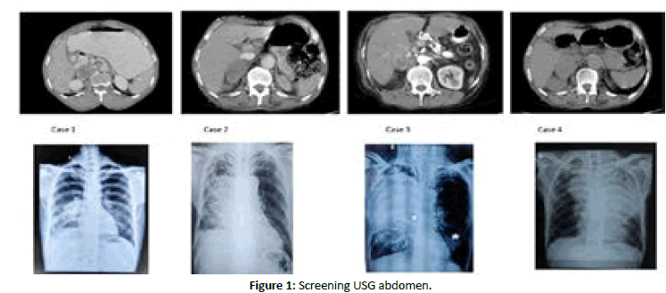
Screening USG was done and showed telescoping of bowel loops at the infra umbilical region with a heterogenous predominantly hypoechoic mass noted over the right suprarenal region, pushing the kidneys medial and posteriorly with minimal vascularity on colour doppler assessment. Overall imaging features were suggestive of right suprarenal metastatic deposit with ileoileal intussusception [6]. The primary imaging was further confirmed with contrast enhanced CT abdomen which showed ileo-ileal intussusception with whirlpool sign and right adrenal metastatic deposit.
Case 2
A 65 years old male came with chief complaints of shortness of breath since three months and chest pain on the right side, cough with expectoration since 3 months with loss of weight and loss of appetite.
A chest X ray PA view was done showed a radio opacity in the right middle zone with silhouetting and multiple air bronchograms without mediastinal shift with blunting of right cp angle. Primary ultrasound screening showed 4.7 × 3.2 cm sized heterogenous predominantly hypoechoic lesion with few echogenic foci within it at the right suprarenal space. Also an irregular ill-defined hyper echoic lesion with minimal vascularity on doppler assessment was noted in segment 6 of liver. CT imaging confirmed USG findings, which showed an enhancing mass lesion in the postero inferior right lobe of liver and infiltrating metastases of the right adrenal gland.
Case 3
A 68 years male, chronic smoker came with complaints of shortness of breath, cough with expectoration for past 3 months and constipation since past 5 days. Patient’s vitals were stable at the time of admission.
Chest X ray showed an ill-defined radio opaque lesion in the right middle zone without mediastinal shift or cp angle blunting. Screening ultrasound of the abdomen showed hypoechoic well defined lesion in the right suprarenal space with minimal vascularity on color doppler [7]. Imaging findings were confirmed with CT abdomen which showed 2.1 × 2.1 cm sized heterogeneously enhancing mass lesion in the right suprarenal space.
Case 4
A 55 years male came with complaints of chest pain, shortness of breath and cough with expectoration for the past 2 months, loss of weight and loss of appetite for 2 months and hemoptysis for 1 and ½ month, chronic smoker. Patient’s vitals were stable at the time of admission.
Chest X ray showed a heterogenous, ill defined radio opacity in the left upper zone of the lung fields with mediastinal shift to the right and hilar opacities noted in the right side. Screening USG abdomen showed a large ill defined heterogenous, predominantly hypoechoic mass lesion in the right suprarenal fossa measuring appx 6.5 × 6.2 cm infiltrating into the psteroinferior segment of right lobe of liver with infiltration also seen in the right kidney upper pole. A similar lesion is also seen in the left suprarenal fossa measuring 3.2 × 3.5 cm. CT imaging showed bilateral adrenal metastatic deposits with primary lung neoplasm. The right adrenal metastatic deposit was seen infiltrating into the upper pole of right kidney with invasion of right renal vein. Biopsy shows squamous cell carcinoma primary lung (Figure 1).

Figure 1: Screening USG abdomen.
Results and Discussion
Lung cancer is one of the important causes of cancer related deaths in our country. Associated adrenaI metastasis can be a deciding factor on patient prognosis depending on the side, functioning status and histological type of metastasis. NSCLC variants spread predominantly to adrenals compared to SCLC type, with poorer prognosis noted in contralateral adrenal mets by hematogenous dissemination.
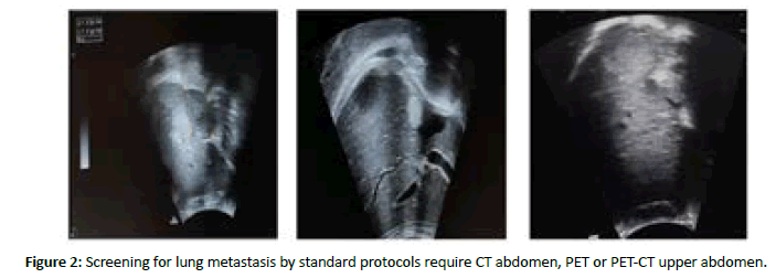
Ultrasound was used as the primary screening modality, considering the cost effectiveness, noninvasive nature and easy availability. A curvilinear 3-6 Mhz probe was used to assess the major organs of upper abdomen using the mindray DC 30 USG Machine and GE [8]. USG findings were further confirmed by CECT Abdomen for all study patients and proven adrenal metastases in all cases. In our case series, we present patients with lung cancer metastasizing to either of the adrenals. All patients are histopathologically proven cases of Ca Lung corresponding to squamous cell carcinoma subtype, sent for screening workup to look for metastases in upper abdomen. Ultrasound was effectively able to detect adrenal metastasis in all cases. The presence of adrenal metastasis in lung cancer equals disseminated disease where patient prognosis is poor, with overall survival rates corresponding to 30 months and a median survival rate of 31%, while an early diagnosis and surgical resection can improve patient outcome by 125 months (Figure 2).

Figure 2: Screening for lung metastasis by standard protocols require CT abdomen, PET or PET-CT upper abdomen.
Screening for lung metastasis by standard protocols require CT abdomen, PET or PET-CT upper abdomen, which are costly, have logistic disadvantages and invasive (requiring contrast administration which has its own complications). Ultrasound is a cheaper and effective alternative (7), with better patient compliance, noninvasive and requires comparatively lesser time for imaging. In our study, ultrasound was effectively able to screen adrenal metastasis in all 4 patients which was confirmed in CT abdomen. Use of Ultrasound as a screening tool for adrenal metastasis in lung cancer patients is a less researched topic, especially in the Indian population. Considering the cost effectives, lesser screening time and noninvasive nature, use of ultrasound can increase identifying adrenal metastasis effectively. Our study lacks in the fact that ultrasound is an operator dependent imaging modality and lesser patients were considered for this study.
Conclusion
Primary adrenal tumors of either cortical or medullary origin have diverse imaging appearances. Using various imaging techniques and correlation with clinical symptoms and functional assays, definitive diagnoses can be given in most lesions. CT is currently the mainstay of imaging of primary adrenal lesions but is well complemented by MRI and functional assays whenever required. Thus, the availability of various imaging techniques has made diagnosis of primary adrenal lesions fairly straight forward.
References
- Mohan A, Garg A, Gupta A, Sahu S, Choudhari C, et al. Clinical profile of lung cancer in North India: A 10 year analysis of 1862 patients from a tertiary care center. Lung India. 2020; 37:190-197.
[Crossref] [Google Scholar] [PubMed]
- Newton PK, Mason J, Bethel K, Bazhenova LA, Nieva J, et al. A stochastic Markov chain model to describe lung cancer growth and metastasis. PLoS One. 2012; 7:34637.
[Crossref] [Google Scholar] [PubMed]
- Bazhenova L, Newton P, Mason J, Bethel K, Nieva J, et al. Adrenal metastases in lung cancer: Clinical implications of a mathematical model. J ThoRac Oncol. 2001; 9:442-446.
[Google Scholar] [PubMed]
- Singh N, Madan K, Aggarwal AN, Das A. Symptomatic large bilateral adrenal metastases at presentation in small cell lung cancer: A case report and review of the literature. J Thorac Dis. 2013; 5:83-86.
[Crossref] [Google Scholar] [PubMed]
- Uemura S, Yasuda I, Kato T, Doi S, Kawaguchi J, et al. Preoperative routine evaluation of bilateral adrenal glands by endoscopic ultrasound and fine-needle aspiration in patients with potentially resectable lung cancer. Endoscopy. 2013; 45:195-201.
[Crossref] [Google Scholar] [PubMed]
- Herth FJ, Eberhardt R, Krasnik M, Ernst A. Endobronchial ultrasound guided transbronchial needle aspiration of lymph nodes in the radiologically and positron emission tomography normal mediastinum in patients with lung cancer. Chest. 2008; 133:887-891.
[Crossref] [Google Scholar] [PubMed]
- Shields TW. Screening, diagnosis, and staging of non-small cell lung cancer and consideration of unusual primary tumors of the lungs. Curr Opin Oncol. 1992; 4:299-307.
[Crossref] [Google Scholar] [PubMed]
- Al Fares HK, Abdullaj S, Gokden N, Menon LP, Menon L. Incidental, solitary and unilateral adrenal metastasis as the initial manifestation of lung adenocarcinoma. Cureus. 2022; 14:32628.
[Crossref] [Google Scholar] [PubMed]


